All About Neurodiagnostic Testing
After an accident, many doctors request a round of neurodiagnostic tests, to determine exactly what the accident victim’s state is and what care they need. Read on to learn more about neurodiagnostic testing, including how it works, who needs it, and what it helps doctors discover.
What Is Neurodiagnostic Testing?
Many accidents cause injury to the brain or nervous system. These injuries are less obvious than a cut or a broken bone, and in some cases even the victim themselves may not be aware of the injury. Unfortunately, this doesn’t mean that the injury has no effects, or that it may not show effects later.
In cases where a doctor expects the potential for neurological damage, they’ll order a round of neurodiagnostic tests. Depending on the nature of the accident and the symptoms (or lack of symptoms) the patient is displaying, a doctor may choose from one or more types of test.
Who Needs Neurodiagnostic Testing?
Not every injury requires neurodiagnostic testing. Symptoms that may cause a doctor to order these tests include, but aren’t limited to:
- trauma to the head or spinal cord
- tingling or numbness
- pain that has no clear physical cause
- headaches
- muscle weakness
- changes in mood or personality
- change in reaction speed or reflexes
- fluctuations in hearing, vision, or other perception
- seizures
- paralysis
Neurological symptoms may be apparent immediately after an accident, or they may take days, weeks, or even months to emerge. The hidden nature of many neurological conditions means many doctors will order tests to rule out problems down the line.
Imaging Tests
Imaging tests include X-rays, CT scans, PET scans, and MRIs. Each of these is a unique test, that returns different types of results.
- X-rays use high-frequency radiation to deliver an image of the patient’s bones. This can be useful for determining the extent of damage to the skull or spinal cord. However, X-rays pass through soft tissue, so they are often less useful for discovering other types of injury. CT scans are X-ray tests used to create three-dimensional cross-section images.
- PET scans, or positron emission tomography scans, use radiation to discover problems with soft tissue. To prepare for a PET scan, a patient drinks a radioactive substance. This radiation spreads throughout the body, and collects in soft tissues. The PET scan itself scans the body for that radiation, and creates an image of the soft tissues which can reveal even small biochemical changes.
- MRI scans use magnets and radio waves to discover anomalies in hard and soft tissue. They’re particularly useful for discovering problems in the brain and spinal cord. However, they may not be an option for patients with pacemakers and other metal implants.
Electrical Impulse Detection
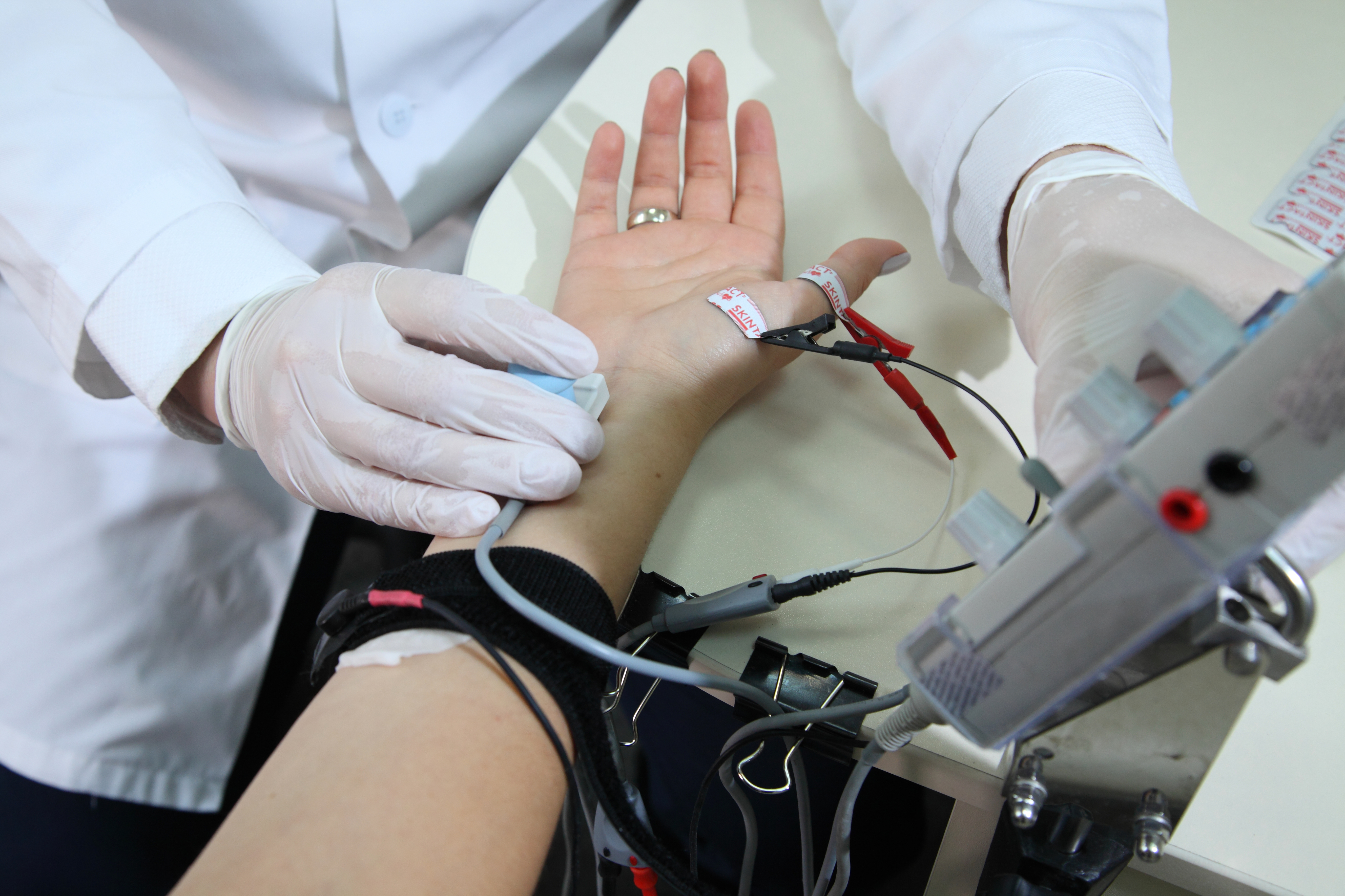
Electrical impulse detection tests include electroencephalograms (EEGs) and electromyographic tests (EMGs). During an EEG, doctors attach small electrode discs to the scalp which monitor brain activity and can reveal problems like seizures, brain damage, or even cerebral hemorrhages.
During an EMG, a doctor inserts a small needle electrode into a muscle and asks a patient to contract the muscle. The test monitors the electrical impulses nerves send to the muscle, and will report any weakness, delayed reactions, or other neurological problems.
Many accidents can result in serious neurological damage, which may not fully appear until some time after the accident. When the doctors at Regional Medical Group perform neurodiagnostic tests, they determine the extent and degree of any damage, and begin devising a treatment plan to help the patient recover.


